Sarcoidosis and Idiopathic Pulmonary Fibrosis: A Diagnostic and Therapeutic Challenge
Received: 20-Jun-2022 / Manuscript No. JCEP-22-63752 / Editor assigned: 23-Jun-2022 / PreQC No. JCEP-22-63752 (PQ) / Reviewed: 11-Jul-2022 / QC No. JCEP-22-63752 / Revised: 20-Jul-2022 / Manuscript No. JCEP-22-63752 (R) / Published Date: 27-Jul-2022 DOI: 10.4172/2161-0681.22.12.410
Abstract
A 59-year-old woman was referred to our Advanced Lung Disease Center for consideration for lung transplantation for stage IV sarcoidosis. She initially presented three years earlier with worsening dyspnea. Highresolution computed tomography of the chest at that time demonstrated ground-glass opacities, traction bronchiectasis, and a 4.5-cm Ascending Aortic Aneurysm (AAA). Biopsy of the lymph nodes revealed noncaseating granulomas. A diagnosis of sarcoidosis with fibrotic changes was plausible; yet, the disease progressed over time despite corticotherapy. At our referral center, a Usual Interstitial Pneumonia (UIP)-Idiopathic Pulmonary Fibrosis (IPF) pattern was observed in the lung parenchyma. The patient was denied transplant due to an AAA requiring risky and complex aortic surgery. Rapid deterioration and refractoriness to corticotherapy suggest her diagnosis may have been sarcoidosis with UIP-IPF pattern. This case illustrates the importance of early recognition of disease patterns of potential transplant candidates and pursuit of interventions to address comorbid conditions in a timely manner.
Keywords: Sarcoidosis; Usual interstitial pneumonia; Idiopathic pulmonary fibrosis; Lung transplantation
Abbreviations
AAA: Ascending Aortic Aneurysm; CSIPF: Combined Sarcoidosis and IPF; HRCT: High Resolution Computed Tomography; IPF: Idiopathic Pulmonary Fibrosis; UIP: Usual Interstitial Pneumonia; VATS: Video-Assisted Thoracoscopy
Introduction
Sarcoidosis and Idiopathic Pulmonary Fibrosis (IPF) are two distinct restrictive lung diseases rarely observed in the same patient, and these pathologies have markedly different clinical and diagnostic features. Few reports have described cases of patients manifesting both sarcoidosis and IPF [1-5], but our understanding of whether these patients present concurrently with two different interstitial lung diseases or with a merging, clinical entity of advanced pulmonary fibrosis is unclear. Differentiating between these diagnoses is a unique challenge for physicians, especially those in the transplant community. The diagnosis is further complicated by the implications it has on prognosis and considerations for treatment. We report an unusual case of sarcoidosis and biopsy-proven Usual Interstitial Pneumonia (UIP)- IPF in an older, Asian woman with a rapid decline in lung function and comorbid cardiovascular conditions.
Case Presentation
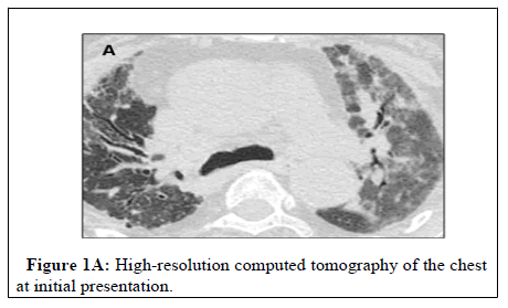
A 59-year-old Asian woman, originally from Bangladesh and living in the United States for 18 years, was referred to our Advanced Lung Disease Center for consideration for lung transplantation for stage IV sarcoidosis. On review of records, the patient had no known prior medical history and had initially presented three years earlier with worsening dyspnea. High Resolution Computed Tomography (HRCT) at that time revealed ground-glass opacities on the left side with interlobular septal thickening and traction bronchiectasis (Figure 1A).
Notably, a 4.5-cm Ascending Aortic Aneurysm (AAA) was also found on concurrent imaging. Infectious causes, including tuberculosis and fungal diseases, were ruled out. Biopsy of the lymph nodes via endobronchial ultrasound-guided transbronchial needle aspiration revealed noncaseating granulomas. Together, these findings supported a diagnosis of sarcoidosis, and the patient was started on oral prednisone (0.3 mg/kg) for 4 weeks, with taper. Her disease was initially responsive to treatment, and short-course corticosteroid bursts were given for every exacerbation. Nonetheless, her lung function progressively declined over time despite corticotherapy, necessitating the referral for evaluation of end-stage lung disease. To our knowledge,treatment with steroid-sparing agents was not employed.
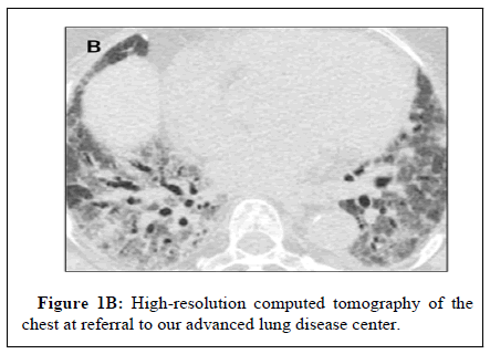
At our center, HRCT of the chest now demonstrated basal groundglass opacities with consolidation in the right lower lobe region and interlobular septal thickening with traction bronchiectasis Figure 1B, consistent with an exacerbation of sarcoidosis.
The patient continued to decline without response to corticosteroid bursts. At this time, she required high-flow oxygen supplementation and physical deconditioning. Histopathological analysis of Video- Assisted Thoracoscopy (VATS)-guided lung tissue biopsy revealed areas of temporal dissociation with honeycombing on lung parenchyma, a typical UIP pattern. Although considered quite atypical of end-stage sarcoidosis, some reports have described end-stage sarcoidosis with the UIP pattern elucidated on histopathological analysis, and we considered this her final diagnosis. This patient was denied evaluation for lung transplant given the AAA (5.9 cm) and risk of performing a complex, open aortic surgery in a patient with endstage lung disease. The patient continued to follow with her pulmonologist at our Advanced Lung Disease Center. Ultimately, she was transitioned into hospice care after worsening disease progression.
Results and Discussion
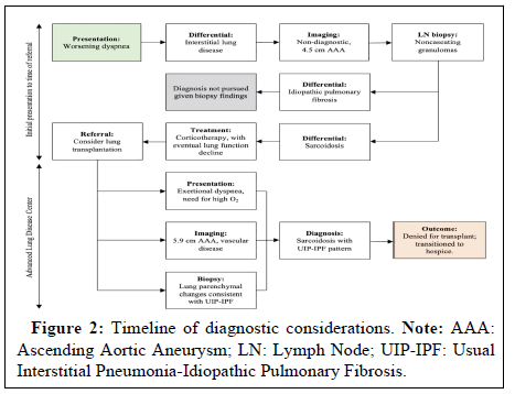
We describe an unusual case of biopsy-proven pulmonary sarcoidosis and UIP-IPF in an older, Asian woman with a rapid decline in lung function and comorbid cardiovascular conditions seeking evaluation for a lung transplant. Our diagnostic considerations are illustrated in Figure 2.
The patient’s initial radiographic findings mimicked the typical radiographic pattern observed in pulmonary sarcoidosis [6]. These findings, combined with her clinical presentation, granulomas on lymph node biopsy, and initial response to corticotherapy (at the referring center), made the diagnosis of sarcoidosis plausible [7-9]. At the time of referral to our center, histopathology of VATS-guided lung tissue biopsy demonstrated parenchymal changes consistent with UIP, which may be only rarely observed in end-stage sarcoidosis. These biopsy findings, rapid deterioration, and refractoriness to corticotherapy suggested that her diagnosis may not have been sarcoidosis, but rather an overlapping disease entity with IPF. Sarcoidosis is also particularly uncommon in this patient’s demographic, as it typically presents in the second through fourth decades of life, and Asians and Hispanics have the lowest rates of incidence, between 3 and 4 per 100,000 [10]. The final diagnosis of end-stage pulmonary sarcoidosis with UIP pattern was not considered until (1) biopsy of lung tissue supported a coincident disease process and (2) the patient failed to respond to corticotherapy at our center, demonstrating the diagnostic challenge presented by this patient as well as the importance of early disease pattern recognition. Therapy may have been better targeted to this patient’s disease process had it been elucidated earlier.
Few reports have previously identified a subset of fibrotic sarcoidosis patients with UIP pattern presenting with rapid clinical deterioration [1-5,11]. Recently, Collins and colleagues characterized this unique presentation in a series of 25 patients with a described “Combined Sarcoidosis and IPF” (CSIPF). Uncertainty with the term CSIPF has since followed, as the most recent diagnostic criteria of IPF requires the exclusion of any other interstitial lung disease [12]. Debate ensues on the proper classification of patients who share radiographic and histopathologic features of sarcoidosis and IPF. It remains unclear whether our case supports a concurrent presentation of two distinct interstitial lung diseases or a merging phenotype of advanced pulmonary fibrosis. Because our patient presented with a plausible diagnosis of sarcoidosis three years prior to demonstrating evidence for IPF, it is unlikely that this patient represents a case of a merging clinical entity (ie, CSIPF). Nonetheless, from the experiences detailed in the literature, it remains possible for patients to develop IPF and sarcoidosis in close proximity, and additional research is warranted to understand whether these patients have a unique genetic predisposition to manifest both diseases.
Conclusion
In conclusion, this patient’s eventual denial of a lung transplant evaluation emphasizes the importance of early recognition of these distinct disease patterns for potential transplant candidates. Interventions to address comorbid conditions, particularly cardiovascular-related, in potential lung transplant candidates should be pursued in a timely manner to reduce disease severity and improve the opportunity for safe listing. Although we recognize that there are limitations in observations from a single case, and this report cannot answer the question of whether the concurrence of sarcoidosis with UIP-IPF pattern represents a combined entity, it is unlikely that this patient represents a case of a merging phenotype given the timeline of her diagnoses.
Source(s) of Support
This manuscript did not receive any specific grant from funding agencies in public, commercial, or not-for-profit sectors.
Conflicting Interest
Authors have no conflicts of interest or financial ties to disclose.
Authorship
All authors have made substantial contributions to all of the following:
• The conception and design of the study, or acquisition of data, or analysis and interpretation of data,
• Drafting the article or revising it critically for important intellectual content,
• Final approval of the version to be submitted.
References
- Collins BF, McClelland RL, Ho LA, Mikacenic CR, Hayes J, et al. (2018) Sarcoidosis and IPF in the same patient-a coincidence, an association or a phenotype?. Respir Med. 144:S20-S27.
[Cross Ref] [Google Scholar] [Pub Med]
- Nobata K, Kasai T, Fujimura M, Mizuguchi M, Nishi K, et al. (2006) Pulmonary sarcoidosis with usual interstitial pneumonia distributed predominantly in the lower lung fields. Intern Med, 45(6):359-362.
[Cross Ref] [Google Scholar] [Pub Med]
- Shigemitsu H, Azuma A (2011) Sarcoidosis and interstitial pulmonary fibrosis; two distinct disorders or two ends of the same spectrum. Curr Opin Pulm Med.17(5):303-307.
[Cross Ref] [Google Scholar] [Pub Med]
- Shigemitsu H, Oblad JM, Sharma OP, Koss MN (2010) Chronic interstitial pneumonitis in end-stage sarcoidosis. Eur Respir J. 35(3): 695-697.
- Zhang C, Chan KM, Schmidt LA, Myers JL (2016) Histopathology of explanted lungs from patients with a diagnosis of pulmonary sarcoidosis. Chest, 149(2): 499-507.
[Cross Ref] [Google Scholar] [Pub Med]
- Nunes H, Brillet PY, Valeyre D, Brauner MW, Wells AU (2007) Imaging in sarcoidosis. Semin Respir Crit Care Med. 28(1): 102-120.
- Patterson KC, Strek ME (2013)Pulmonary fibrosis in sarcoidosis. Clinical features and outcomes. Ann Am Thorac Soc. 10(4):362-370.
[Cross Ref] [Google Scholar] [Pub Med]
- Bonham CA, Strek ME, Patterson KC (2016) From granuloma to fibrosis: sarcoidosis associated pulmonary fibrosis. Curr Opin Pulm Med. 22(5):484-491.
[Cross Ref] [Google Scholar] [Pub Med]
- Teirstein AT, Morgenthau AS (2009) "End-stage" pulmonary fibrosis in sarcoidosis. Mt Sinai J Med. 76(1):30-36.
[Cross Ref] [Google Scholar] [Pub Med]
- Arkema EV, Cozier YC (2018) Epidemiology of sarcoidosis: current findings and future directions. Ther Adv Chronic Dis. 9(11):227-240.
[Cross Ref] [Google Scholar] [Pub Med]
- Bianchi F, Piccioli C, Rosi E, Carobene L, Spina D, et al. (2019) Combined sarcoidosis and idiopathic pulmonary fibrosis (CSIPF): A novel disease phenotype?. Respir Med. 160:105650.
[Cross Ref] [Google Scholar] [Pub Med]
- Raghu G, Remy-Jardin M, Myers JL. (2018) Diagnosis of Idiopathic Pulmonary Fibrosis. An Official ATS/ERS/JRS/ALAT Clinical Practice Guideline. Am J Respir Crit Care Med.198(5):e44-e68.
[Cross Ref] [Google Scholar] [Pub Med]
Citation: Olson MT, Roy SB, Walia R, Arjuna A (2022) Sarcoidosis and Idiopathic Pulmonary Fibrosis: A Diagnostic and Therapeutic Challenge. J Clin Exp Pathol. 12: 410. DOI: 10.4172/2161-0681.22.12.410
Copyright: © 2022 Olson MT, et al. This is an open-access article distributed under the terms of the Creative Commons Attribution License, which permits unrestricted use, distribution, and reproduction in any medium, provided the original author and source are credited.
Share This Article
Open Access Journals
Article Tools
Article Usage
- Total views: 1402
- [From(publication date): 0-2022 - Dec 06, 2023]
- Breakdown by view type
- HTML page views: 1179
- PDF downloads: 223


